格物
痛觉感受器1906
nociceptor
sensory neuron that detects pain
A nociceptor is a sensory neuron that responds to damaging or potentially damaging stimuli by sending "possible threat" signals to the spinal cord and the brain. The brain creates the sensation of pain to direct attention to the body part, so the threat can be mitigated; this process is called nociception.
摘自英文维基百科「Nociceptor」(英文维基百科,暂无中文条目),CC BY-SA 4.0
- 发现 / 发明时间
- 1906 年
- 发现者 / 提出者
- 查尔斯·斯科特·谢灵顿
- 学科
- 生物学
- 上级概念
- 感觉细胞
相关科学家

百科正文
本条目暂无中文维基百科条目,下面是英文维基百科原文。
Terminology
Nociception and pain are usually evoked only by pressures and temperatures that are potentially damaging to tissues. This barrier or threshold contrasts with the more sensitive visual, auditory, olfactory, taste, and somatosensory responses to stimuli. The experience of pain is individualistic and can be suppressed by stress or exacerbated by anticipation. Simple activation of a nociceptor does not always lead to perceived pain, because the latter also depends on the frequency of the action potentials, integration of pre- and postsynaptic signals, and influences from higher or central processes.
History
Nociceptors were discovered by Charles Scott Sherrington in 1906. In earlier centuries, scientists believed that animals were like mechanical devices that transformed the energy of sensory stimuli into motor responses. Sherrington's experiments demonstrated that different types of stimulation to an afferent nerve fiber's receptive field led to different responses. Some intense stimuli trigger reflex withdrawal, certain autonomic responses, and pain. The receptors for these stimuli were called nociceptors.
Studies of nociceptors have been conducted on conscious humans as well as surrogate animal models.
Location
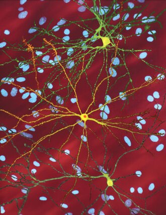
In mammals, nociceptors are found in any area of the body that can sense noxious stimuli. External nociceptors are found in tissue such as the skin (cutaneous nociceptors), the corneas, and the mucosa. Internal nociceptors are found in a variety of organs, such as the muscles, the joints, the bladder, the visceral organs, and the digestive tract. The cell bodies of these neurons are located in either the dorsal root ganglia or the trigeminal ganglia. The trigeminal ganglia are specialized nerves for the face, whereas the dorsal root ganglia are associated with the rest of the body. The axons extend into the peripheral nervous system and terminate in branches to form receptive fields.
Types and functions
Nociceptors are usually electrically silent when not stimulated. The peripheral terminal of the mature nociceptor is where the noxious stimuli are detected and transduced into electrical energy. When the electrical energy reaches a threshold value, an action potential is induced and driven towards the central nervous system (CNS). This leads to the train of events that allows for the conscious awareness of pain. The sensory specificity of nociceptors is established by the high threshold only to particular features of stimuli. Only when the threshold has been reached are the nociceptors triggered.
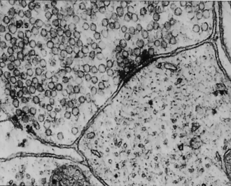
In terms of conduction velocity, nociceptors come in two groups. Aδ fiber axons are myelinated and can allow an action potential to travel towards the CNS at speeds from 5 to 30 meters/second.
Pathways
Nociceptive pain pathways involve the transmission of sensory information from peripheral nociceptors of C-fibers to the dorsal horn of the spinal cord. These synaptic signals are transmitted to second-order neurons in laminae I and II, which serve as initiation sites for nociceptive information entering the central nervous system. These neurons play a crucial role in regulating the degree of pain transmission that crosses to the contralateral side and ascends to the primary tract, where the spinothalamic and spinoreticular pathways are. These ascending spinal cord pathways convey and localize nociceptive pain information from the periphery (body) to the brainstem.
Sensitivity
Nociceptor sensitivity is modulated by a large variety of mediators in the extracellular space, such as toxic and inflammatory molecules. Peripheral sensitization represents a form of functional plasticity of the nociceptor. The nociceptor can change from being simply a noxious stimulus detector to a detector of non-noxious stimuli. The result is that low intensity stimuli from regular activity initiates a painful sensation. This is commonly known as hyperalgesia. Inflammation is one common cause that results in the sensitization of nociceptors. Normally hyperalgesia ceases when inflammation goes down, however, sometimes genetic defects and/or repeated injury can result in allodynia: a completely non-noxious stimulus like light touch causes extreme pain. Allodynia can also be caused when a nociceptor is damaged in the peripheral nerves.
Neural development
Nociceptors develop from neural-crest stem cells during embryogenesis. The neural crest is responsible for a large part of early development in vertebrates. It is specifically responsible for development of the peripheral nervous system (PNS). The neural-crest stem cells split from the neural tube as it closes, and nociceptors grow from the dorsal part of this neural-crest tissue. They form late during neurogenesis. Earlier forming cells from this region can become non-pain sensing receptors, either proprioceptors or low-threshold mechanoreceptors. All neurons derived from the neural crest, including embryonic nociceptors, express the tropomyosin receptor kinase A (TrkA), which is a receptor to nerve growth factor (NGF). However, transcription factors that determine the type of nociceptor remain unclear.
In other animals
Nociception has been documented in non-mammalian animals, including fish and a wide range of invertebrates, including leeches, nematode worms, sea slugs, and larval fruit flies. Although these neurons may have pathways and relationships to the central nervous system that are different from those of mammalian nociceptors, nociceptive neurons in non-mammals often fire in response to similar stimuli as mammals, such as high temperature (40 degrees C or more), low pH, capsaicin, and tissue damage.
For example, in fruit flies, specific multidendritic sensory neurons play a role in nociception. In mollusks, nociceptive responses are mediated by pedal sensory neurons.
以上节选摘自英文维基百科「Nociceptor」,按 CC BY-SA 4.0 使用,去掉了参考文献、外部链接等尾部小节,公式改写成了纯文本。篇幅所限,较长的小节只摘了开头一部分,有的小节没有收;全文、配图和出处见原文。